Small intestines rock! And finally get some well-deserved respect
By Melanie Medina
To get a good look at our digestive system, gastroenterologists can go in from the top with an endoscopy,
or from the bottom with a colonoscopy.
But what about the middle — several feet located in our small intestine? This area is a little trickier
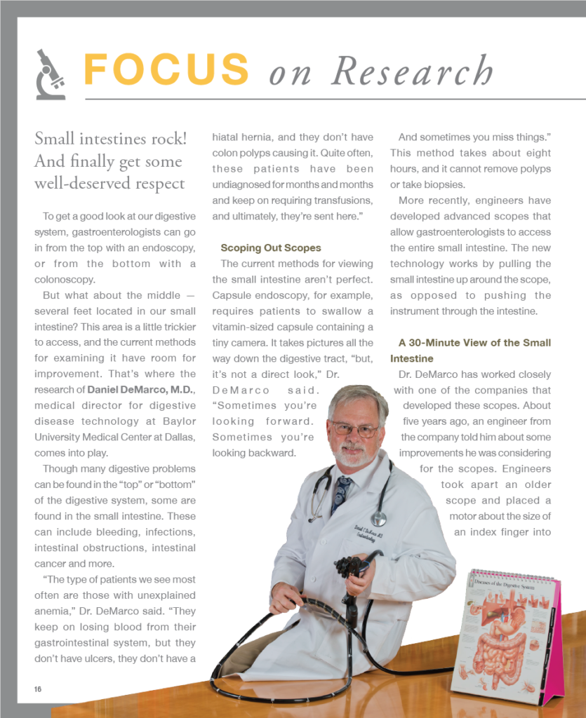
to access, and the current methods for examining it have room for improvement. That’s where the research of Daniel DeMarco, M.D., medical director for digestive disease technology at Baylor University Medical Center at Dallas, comes into play.
Though many digestive problems can be found in the “top” or “bottom” of the digestive system, some are
found in the small intestine. These can include bleeding, infections, intestinal obstructions, intestinal cancer and more.
“The type of patients we see most often are those with unexplained anemia,” Dr. DeMarco said. “They keep on losing blood from their gastrointestinal system, but they don’t have ulcers, they don’t have a hiatal hernia, and they don’t have colon polyps causing it. Quite often, these patients have been undiagnosed for months and months and keep on requiring transfusions,
and ultimately, they’re sent here.”
Scoping Out Scopes
The current methods for viewing the small intestine aren’t perfect. Capsule endoscopy, for example, requires patients to swallow a vitamin-sized capsule containing a tiny camera. It takes pictures all the way down the digestive tract, “but,
it’s not a direct look,” Dr. DeMarco said.
“Sometimes you’re looking forward. Sometimes you’re looking backward. And sometimes you miss things.” This method takes about eight hours, and it cannot remove polyps or take biopsies.
More recently, engineers have developed advanced scopes that allow gastroenterologists to access the entire small intestine. The new technology works by pulling the small intestine up around the scope, as opposed to pushing the instrument through the intestine.
A 30-Minute View of the Small Intestine
Dr. DeMarco has worked closely with one of the companies that developed these scopes. About five years ago, an engineer from the company told him about some improvements he was considering for the scopes. Engineers took apart an older scope and placed a motor about the size of an index finger into the handle. The motor turns a cable that runs down inside the scope to a bushing on the end of the scope turning a spiral segment.
“The cable inside the scope is kind of like a speedometer cable,” Dr. DeMarco said. After several iterations, Olympus, the company that now makes the scope, submitted it to the Food and Drug Administration, where it received preliminary approval (a step before it is fully FDA approved). The Baylor Research Institute Institutional Review Board (IRB) has approved its use for a research trial here and earlier this year, Dr. DeMarco began using the scope on patients.
About six patients have undergone endoscopy with the scope, and Dr. DeMarco expects that number to triple by the end of the year. Two other centers — the University of Massachusetts and the University of Florida in Gainesville — are also enrolling patients in the trial. “The company’s next step is to get this approved by the FDA,” Dr. DeMarco said. “And then get it manufactured and have one in every major medical center.
This article was originally published in the Winter 2016 issue of The Torch, a quarterly publication from the Baylor Scott & White Health Foundation-Dallas.


Leave a Comment: